Prominent Ear Aesthetics
EBOPRAS-certified plastic surgeon Op. Dr. Necdet Urhan performs otoplasty (prominent ear correction) in Antalya using the Mustardé suture technique for antihelix reconstruction and the Furnas conchal setback technique, bringing the cranioauricular angle to the ideal 25–30° range through a hidden retroauricular incision; results are permanent and the scar remains invisible from the front.
"Prominent ear deformity rarely involves a single anatomical issue. In most patients, antihelix underdevelopment, conchal hypertrophy, and lobule position must each be assessed independently — and the surgical technique must be individually tailored. A one-size-fits-all approach significantly increases the risk of post-operative asymmetry and relapse."
— Op. Dr. Necdet Urhan, Specialist in Plastic and Aesthetic Surgery, Antalya
Medical Disclaimer: This page is for general informational purposes only and does not constitute medical advice. The decision to undergo otoplasty requires a personalised consultation and professional evaluation.
What Is Prominent Ear? Anatomical Causes
Prominent ear (protruding ear, bat ear, or otapostasis) is an aesthetic deformity in which the ears project markedly away from the side of the head. Because the majority of ear development is completed before birth and during the first two years of life, this structural condition is almost always congenital and stems from specific cartilage deficiencies.
In a normally positioned ear, the cranioauricular angle — the angle between the plane of the auricle and the scalp — measures 25–30°. In prominent ear, this angle exceeds 35°, making the ears conspicuous both in profile and from the front. According to Op. Dr. Necdet Urhan, the majority of prominent ear cases involve more than one anatomical deficit simultaneously, and treatment must therefore be individually planned rather than protocol-driven.
Key Anatomical Structures Involved
Antihelix Underdevelopment
The antihelix is the inner curved ridge running parallel to the outer helical rim. When it fails to fold adequately during development, the upper ear rotates outward. This is the most common cause of prominent ear and is addressed with the Mustardé suture technique.
Conchal Hypertrophy
The concha — the bowl-shaped central depression of the ear (cymba concha + cavum concha) — pushes the ear forward when overdeveloped. The Furnas conchal setback technique approximates the concha to the mastoid fascia to reduce protrusion.
Scapha Width
The scapha is the flat triangular region between the antihelix and the helical rim. When excessively wide and flat, it contributes to the overall flattened appearance of the ear. Cartilage scoring along the scapha can help create the antihelix fold.
Helical Rim Position
The outer boundary of the ear — the helical rim — can travel in a direction that causes the upper pole of the ear to protrude further than the lower. Upper pole prominence is evaluated separately in surgical planning and requires specific correction.
Lobule Position
The ear lobe (lobulus auriculae) may protrude downward or outward independently of the cartilaginous framework. After cartilage correction, a visible lobule mismatch can create residual asymmetry; lobule reduction is applied when needed.
Cranioauricular Angle
This is the angle the ear makes relative to the scalp surface. Normal: 25–30°. Prominent ear: above 35°. The surgical goal is to bring both ears symmetrically within the normal range — not to pin them flat against the head, which creates an over-corrected, unnatural look.
Surgical Techniques
Op. Dr. Necdet Urhan selects and combines techniques based on each patient's specific anatomical findings. All incisions are placed in the retroauricular sulcus — the natural crease behind the ear — making scarring essentially invisible.
Technique Comparison
| Technique | Target Structure | Cartilage Removal | Best Indication |
|---|---|---|---|
| Mustardé | Antihelix | No | Antihelix underdevelopment |
| Furnas | Concha | No | Conchal hypertrophy |
| Scoring / Abrading | Scapha / Cartilage | Partial | Rigid cartilage |
| Mustardé + Furnas | Antihelix + Concha | No | Combined deformity (most common) |
Surgical Process: 5 Stages
Stage 1 — Evaluation and Planning
The cranioauricular angle, antihelix morphology, conchal depth, lobule position, and cartilage flexibility are each assessed individually. The patient's age and cartilage resilience inform technique selection. Photographic analysis establishes the target angle and symmetry goals.
Stage 2 — Anaesthesia
In adults and older children, local anaesthesia with sedation provides a comfortable experience. In children under 6, general anaesthesia is preferred. Op. Dr. Necdet Urhan establishes the anaesthesia plan individually for each patient.
Stage 3 — Incision and Cartilage Access
A 1–2 cm incision is made in the retroauricular sulcus — the natural crease behind the ear. The skin and subcutaneous tissue are gently elevated to expose the posterior cartilage surface. Vascular pedicles are preserved to protect skin viability.
Stage 4 — Cartilage Reshaping and Fixation
The individually selected technique combination is applied: Mustardé sutures recreate the antihelix fold; Furnas sutures approximate the concha to the mastoid fascia when needed. Intraoperative symmetry is checked by sitting the patient up or viewing the ear in the natural head position.
Stage 5 — Closure and Dressing
The incision is closed with 4-0 or 5-0 absorbable sutures. Cotton bolsters are placed between the ear and the scalp to maintain contour, and a compressive head dressing is applied. The dressing remains in place for 5–7 days; a soft headband is then used for 2–3 further weeks.
Ideal Candidate Criteria
Age: Children 6+, Adults Any Age
Ear cartilage reaches approximately 90% of adult size by age 5–6. Surgery is therefore planned from age 6 onwards. For adults, there is no upper age limit — cartilage flexibility decreases with age but does not preclude surgery.
Aesthetic Concern or Psychological Impact
Individuals who feel self-conscious, avoid social situations, or restrict their hairstyle choices due to prominent ears are excellent candidates. In children, early correction before school age can prevent peer-related psychological pressure.
Overall Health
Chronic conditions should be well-controlled; active smoking should be discontinued prior to surgery; blood-thinning medications should be stopped in consultation with the prescribing physician. Routine pre-operative health assessment is performed.
Realistic Expectations
The aim is natural, proportionate ear positioning relative to the face — not perfect symmetry. Minor natural asymmetry between the two ears is a normal anatomical variation and is discussed openly during consultation.
Post-operative Compliance
Willingness to wear the compressive headband and follow post-operative instructions for 4–6 weeks is essential. In children, parental cooperation with the recovery protocol significantly improves long-term outcomes.
Contraindications
Active ear infection or skin disease, uncontrolled diabetes, known keloid formation tendency, or active connective tissue disorders are factors assessed at consultation. These may require optimisation or alternative planning.
Advantages of Otoplasty
Permanent, Natural Result
Once the cartilage settles into its new position (3–6 months post-operatively), results are permanent. As the cartilage fully integrates, the outcome becomes increasingly natural — no "operated" appearance.
Hidden Scar
The retroauricular incision is completely concealed from the front. The scar is invisible during normal social interaction and fades to near-invisibility within 12–18 months.
Rapid Social Recovery
Most patients return to desk work and social activities within 5–7 days. Adults can conceal the ears under their hair during the early recovery period. Children can typically return to school within a week.
Hearing Is Unaffected
Otoplasty involves only the external ear cartilage (pinna). The middle ear, inner ear, and auditory nerve are completely untouched. Hearing function is not affected in any way.
Psychological Benefit in Childhood
Early correction eliminates a common source of teasing and social anxiety during school years, making a meaningful contribution to the child's confidence and social development at a formative stage.
Hairstyle Freedom
Buns, ponytails, short cuts, and other styles that expose the ears — long avoided due to self-consciousness — become freely accessible choices after surgery.
Recovery Timeline and Care
| Period | What to Expect and Recommendations |
|---|---|
| Days 1–3 | The compressive head dressing remains in place. Mild to moderate pain and a feeling of tightness are normal; managed with prescribed analgesics. Sleeping with the head elevated is recommended. |
| Days 5–7 | Follow-up visit: the bandage is removed, sutures are assessed, and ear position is evaluated. Most adults can return to desk-based work. Children can typically return to school with precautions. |
| Weeks 2–3 | A soft headband is worn nightly to maintain ear position during sleep — critical for long-term result stability. Mild swelling and skin discolouration in this period are normal and expected. |
| Weeks 4–6 | Contact sports and swimming must be avoided. Direct sun exposure to the scar area should be minimised; SPF 50+ sunscreen is recommended. Exercise may be gradually resumed. |
| Months 3–6 | Final results become fully apparent as the cartilage completes its remodelling process. The scar continues to soften and fade. All sports and activities may be fully resumed. |
Important: Full compliance with the headband protocol is the single most important patient-controlled factor in preventing relapse. According to Op. Dr. Necdet Urhan, consistent headband use during the first 6 weeks — especially during sleep — is what separates excellent from average long-term outcomes, particularly in paediatric patients.
Combination Procedures
Otoplasty can be planned as a standalone procedure or combined with other facial aesthetic surgeries. According to Op. Dr. Necdet Urhan, facial proportions should be assessed as a whole — ear position, nose shape, chin projection, and overall facial balance are interrelated and benefit from coordinated planning.
Rhinoplasty
Nose shape and ear position together define the overall facial profile. Combined planning allows for a single anaesthesia and a unified recovery period. Rhinoplasty →
Eyelid Aesthetics (Blepharoplasty)
Upper and lower eyelid appearance is assessed as part of the broader facial harmony evaluation alongside ear position. Eyelid Aesthetics →
Submental Liposuction
In patients concerned with both ear prominence and jaw-neck definition, submental liposuction may be combined with otoplasty to refine the overall facial profile. Submental Liposuction →
Chin Shaping (Chinoplasty)
Chin projection and ear position together define the lateral facial profile. Combined assessment and correction may benefit patients seeking comprehensive facial balance. Chin Shaping →
Cost and Pricing
The cost of otoplasty varies depending on the complexity of the technique required (single vs. combined), whether correction is unilateral or bilateral, the type of anaesthesia, and any additional adjunct procedures. At Op. Dr. Necdet Urhan's clinic, pricing is presented transparently following the consultation examination.
Factors Affecting Price:
- Unilateral or bilateral procedure
- Single technique (Mustardé or Furnas) vs. combined approach
- Local anaesthesia with sedation vs. general anaesthesia
- Need for adjunct lobule reduction
- Any additional combined facial procedures
Antalya offers internationally competitive pricing for otoplasty compared to Western Europe and North America, making it a preferred destination for medical tourists. For detailed pricing, please schedule a consultation. Explore facelift procedures →
Why Op. Dr. Necdet Urhan?
EBOPRAS Certification
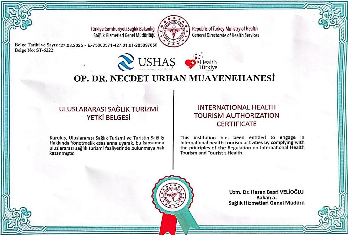
Certification from the European Board of Plastic, Reconstructive and Aesthetic Surgery (EBOPRAS) confirms that Op. Dr. Necdet Urhan has met the rigorous European standards for training, knowledge, and clinical competence in plastic surgery.
ISAPS Membership
Membership in the International Society of Aesthetic Plastic Surgery (ISAPS) reflects ongoing commitment to the latest evidence-based techniques and international ethical standards in aesthetic practice.
Focused on Facial Aesthetics
Op. Dr. Necdet Urhan focuses exclusively on facial aesthetics. This concentrated specialisation means cumulative expertise in ear, nose, eyelid, and facial harmony procedures — with ear correction as a core component of this focused practice.
Individualised Surgical Plan
No two ears are anatomically identical. Op. Dr. Urhan devises a case-specific technique combination rather than applying a standard protocol, reducing the risk of relapse, telephone deformity, and asymmetry.
International Patient Experience
Located in Muratpaşa, Antalya, the clinic regularly receives patients from the UK, Germany, Scandinavia, and beyond. Multilingual support, accommodation coordination, and video follow-up consultations are available.
Structured Follow-up Care
Post-operative visits are scheduled systematically to monitor healing, assess scar maturation, and ensure that cartilage position remains stable. Patient safety and long-term satisfaction are the primary clinical goals.
Risks and Complications
Otoplasty is generally a safe procedure, but as with all surgery, specific risks exist. According to Op. Dr. Necdet Urhan, the vast majority of cases proceed without complications; meticulous patient selection and surgical technique minimise the risk profile substantially.
Asymmetry
The most common reported concern. Minor asymmetry is a normal anatomical variation; noticeable asymmetry requiring revision occurs in a small proportion of cases and is addressed at follow-up.
Relapse
If sutures loosen before full cartilage remodelling is complete, the ear may partially return to its original position. Correct suture selection and headband compliance are the main preventive factors.
Haematoma / Seroma
Blood or serous fluid collection behind the ear may occur within the first 48–72 hours. The compressive dressing reduces this risk; drainage is performed if it develops.
Temporary Sensation Changes
Tingling, numbness, or altered sensation in the upper ear may persist for several weeks to months post-operatively and typically resolves spontaneously as local nerve endings recover.
Infection / Chondritis
Pre- and post-operative antibiotic prophylaxis minimises infection risk. Cartilage infection (chondritis), while rare, is a serious complication requiring prompt antibiotic treatment and occasionally surgical drainage.
Telephone Deformity
Over-correction of the upper and lower poles with under-correction of the middle creates an appearance resembling a telephone handset. This is a technique-related issue prevented by careful suture tension calibration.
Frequently Asked Questions
Book a Consultation for Otoplasty in Antalya
Schedule your personal evaluation with Op. Dr. Necdet Urhan. At consultation, your antihelix morphology, conchal depth, and the optimal technique combination will be assessed and explained in full.
📍 Muratpaşa, Antalya | EBOPRAS Certified | ISAPS Member